| Mycobacteriosis in Fish | ||||||
| Page 2 of 5 | Pages: 1 . 2 . 3 . 4 . 5 | |||||
Signs of Mycobacteriosis in Fish Because of the slow progression of the disease, younger fish infected with mycobacteriosis show no external signs. As fish age or are stressed, the infection becomes more serious. A chronic progressive disease develops that may be characterized by emaciation, inflammation of the skin, exophthalmia ("pop-eye"), open lesions, or ulceration. Fish may become sluggish and bloated, refuse to eat, and develop fin and tail rot as well as scale loss. Internally, gray-white granulomas (nodules) develop in the liver, kidney, spleen, heart, and muscles ( Figure 1 ). When the nodules develop in the organs, edema (excess fluid accumulation) may develop, as well as peritonitis (inflammation of the body cavity). If infection spreads to the skeletal system, deformities such as a bent spine may be noticed. Eventually, infected fish succumb and die. 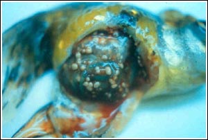 Figure 1 Granulomas (nodules) are readily visible on multiple organs in the abdominal cavity during necropsy. The disease in this fish was very advanced.
Diagnosis A presumptive diagnosis of mycobacteriosis can be based on the presence of typical granulomas, which will be seen at necropsy (Figure 1). The fish shown in Figure 1 has a very advanced infection. However, it is possible to see granulomas using a light microscope by making wet mounts of organ tissue ( Figure 2 ). For this reason, a wet mount should be made of liver, spleen, and kidney tissue as part of a routine necropsy. Granulomas will also often be present on skin ulcerations. If granulomas are seen, an acid-fast stain should be performed immediately on suspect tissue. In the laboratory, granulomatous material is smeared onto a slide, dried, heat-fixed, and stained with Kinyoun or Ziehl-Nielsen stain to confirm the presence of acid-fast bacteria. A positive acid-fast stain reveals red to pink rod-shaped bacteria against a light green background. The bacteria will appear in the center of, or surrounding suspect lesions. A positive acid-fast stain very strongly supports the diagnosis. A negative acid-fast stain (no red or pink rods seen) suggests another cause for the granulomas, such as parasites or fungus. 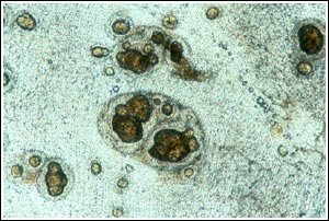 Figure 2: Granulomas in an unstained wet mount. When this tissue change is observed, an acid-fast stain is indicated.
Fish suspected of being infected with mycobacteria should be submitted to a diagnostic laboratory to confirm the infection, and, if possible, to culture and identify the causative agent. Acid-stains will be performed on fresh tissue as described above, in addition to acid-fast stains of histopathological samples. Crushed granulomas are also streaked onto Lowenstein-Jensen or Dorset media, specialized growth media for mycobacteria, where growth can take up to 28 days. Confirmation of the diagnosis by culture of the organism is desirable, although no growth does not necessarily rule out mycobacteria, especially if acid-fast organisms are present. more ... |
 |
|||||
| About Us :: Message Board :: Chat | |||||
| Library :: Photo Gallery :: Links & Resources :: Breeders & Sponsors :: Merchandise | |||||
| Website designed by: EthanCote.com | © 2001-2004, SimplyDiscus.com. All Rights Reserved. | ||||